
FIG 7.
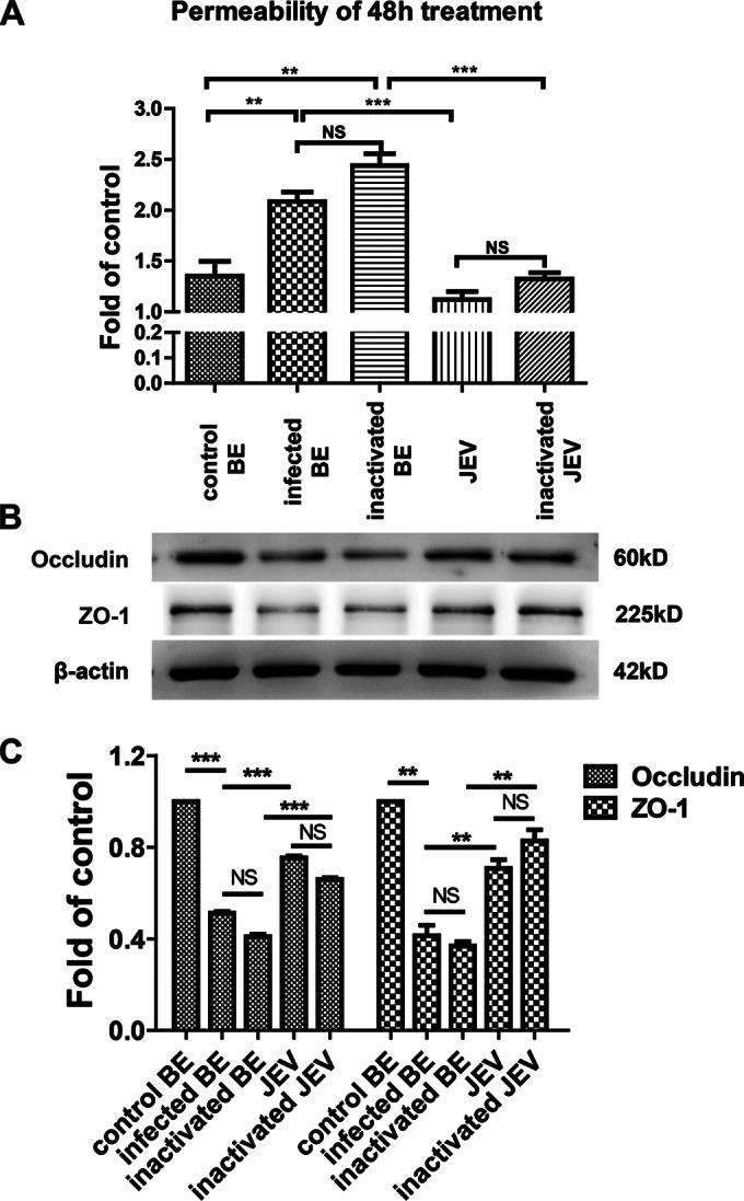
Effects of brain extracts on endothelial barrier permeability. (A) After b.End3 monolayers were grown to confluence on transwell membranes, the cells were treated with brain extracts, UV-inactivated brain extracts, JEV, or UV-inactivated JEV for 24 h or 48 h. FITC-dextran-10000 was applied apically at 1 mg/ml for 30 min, and then the permeability was measured from the samples of lower chamber with a fluorometer (excitation, 492 nm; emission, 520 nm). (B) Mouse BMECs were treated as described above, and then protein samples were collected and subjected to Western blotting for the TJ proteins occludin, claudin-5, and ZO-1. β-Actin was used as a loading control. (C) The results of panel B were normalized with β-actin and densimetrically quantified as the fold change versus control brain extracts (control BE). Means ± SEM of three independent experiments were shown. Asterisks indicate statistical significance (*, P < 0.05; **, P < 0.01; ***, P < 0.005).