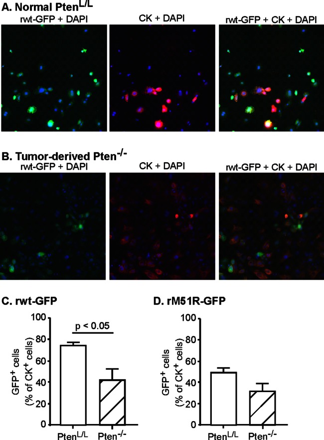
FIG 2.
Prostate tumors contain a mixture of resistant and susceptible cells. Primary cultures of prostate epithelial cells were established from Pten−/− prostate tumors of 3-month-old mice and their littermate normal PtenL/L controls. Cells were seeded into imaging plates and were infected with rwt-GFP or rM51R-GFP viruses (green). Cells were fixed, permeabilized, immunolabeled for cytokeratin (red) and stained with DAPI to label cellular DNA (blue), and then analyzed with a high-content imaging system. Representative fluorescence images are shown in panels A and B. The numbers of cells that were positive for GFP and cytokeratin were expressed as a percentage of total cytokeratin-positive cells (C and D). The data shown are averages of at least three experiments ± the standard errors of the mean (SEM).

