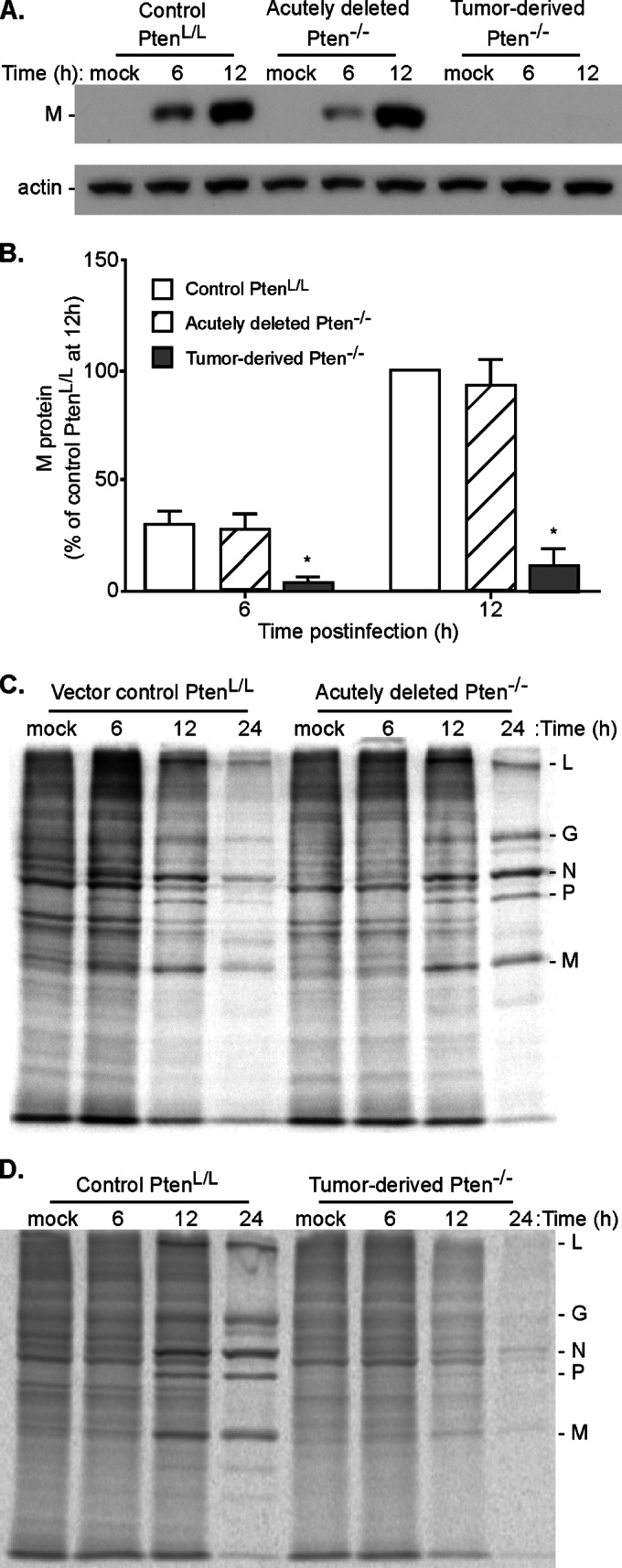
FIG 5.

Viral protein expression in control PtenL/L cells and acutely deleted Pten−/− cells versus tumor-derived Pten−/− cells. (A) Control PtenL/L, acutely deleted Pten−/−, and tumor-derived Pten−/− cells were infected with rwt virus at an MOI of 10. Cell lysates were harvested at 6 and 12 h postinfection and probed for viral matrix protein (M) protein expression by immunoblotting. Representative immunoblots of at least three experiments are shown. (B) Viral matrix (M) protein expression was quantified and expressed as a percentage of M protein expression in control PtenL/L cells at 12 h postinfection. *, P < 0.05. (C) Vector control PtenL/L and acutely deleted Pten−/− cells were infected with rwt virus at an MOI of 10 or mock infected (M). At 6, 12, and 24 h postinfection, the cells were pulse-labeled with [35S]methionine. Cell lysates were harvested and resolved on SDS-PAGE gels. Representative phosphorimages are shown. Viral proteins L, G, N, P, and M are indicated on the right. (D) Nontransduced control PtenL/L and tumor-derived Pten−/− cells were infected with rwt virus at an MOI of 10 or mock infected (M). At 6, 12, and 24 h postinfection, cells were pulse-labeled with [35S]methionine and analyzed as in panel C.
