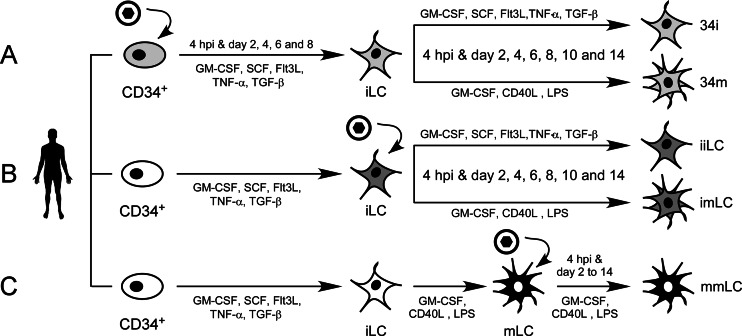
FIG 1.
Schematic representation of the methods used to generate the five different types of dendritic cells analyzed in this work. Umbilical cord blood-derived CD34+ hematopoietic progenitor cells from single donors were partitioned into two groups immediately after thawing. One set was left uninfected (B, white oval), while the other was exposed to TB40-BAC4 or AD169varATCC virions for 4 h (A, light gray oval). Infected and uninfected CD34+ cells were then cultured in iLC differentiation medium for 8 days, with supernatants and cells collected every 2 days. (A) Immature LC derived from infected progenitors were harvested on day eight and replated in either fresh iLC differentiation medium (34i) or maturation medium (34m) for 14 days, with collection of cells and supernatants every 2 days. (B) Immature LC derived from uninfected progenitors were harvested on day eight and exposed to TB40-BAC4 or AD169varATCC virions for 4 h prior to culture in iLC differentiation medium (iiLC) or in maturation medium (imLC) for 14 days, with collection of cells and supernatants every 2 days. (C) A portion of the iLC differentiated from uninfected CD34+ cells was cultured in maturation medium for 2 days to generate mLC, which were then exposed to TB40-BAC4 or AD169varATCC virions for 4 h prior to culture in maturation medium (mmLC) for 14 days, with supernatants and cells collected every 2 days. White ovals and star-shaped outlines represent uninfected progenitor and dendritic cells, respectively, while light gray, dark gray, and black outlines represent infected cells. Black circles enclosing a black hexagon depict CMV virions. GM-CSF, granulocyte-macrophage-colony-stimulating factor; SCF, stem cell factor; Flt3L, Flt3 ligand; TNF-α, tumor necrosis factor alpha; TGF-β, transforming growth factor β1; CD40L, CD40 ligand; LPS, lipopolysaccharide.

