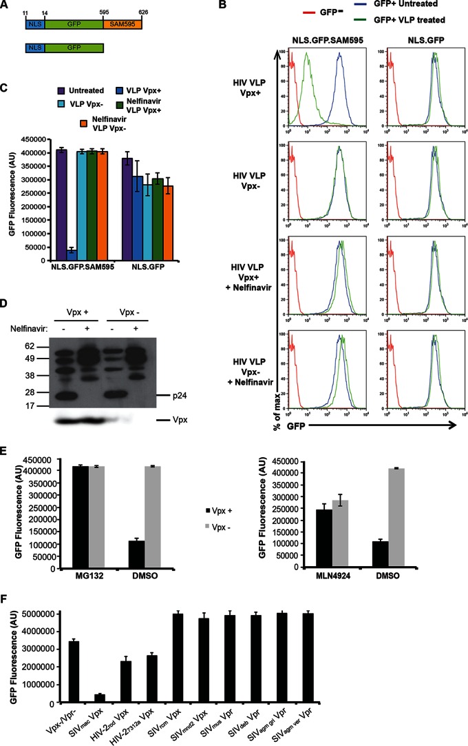
FIG 1.
Protease inhibitor prevents Vpx-induced degradation. (A) The structure of GFP.SAMHD1 fusion proteins used to generate HeLa stable cell lines is diagrammed. The fusion proteins were expressed in lentiviral vectors in which an amino-terminal NLS (KRPR) is fused to GFP with or without the Vpx-binding domain of SAMHD1 (NLS.GFP.SAM595 or NLS.GFP, respectively). Numbers in the diagram indicate the corresponding SAMHD1 amino acids. (B) NLS.GFP.SAM595 and NLS.GFP cell lines were treated with Vpx-containing (Vpx+) or control (Vpx−) VLPs and, after 24 h, analyzed by flow cytometry. As shown in the bottom two rows, the VLPs were produced in cells treated with 3 μM nelfinavir protease inhibitor. (C) At 24 h postinfection, GFP fluorescence of the cells treated as described for panel B was measured on a microplate fluorometer. The bars represent the means of the results determined with quadruplicate wells, and the error bars represent the standard deviations. AU, arbitrary units. (D) VLPs produced in the presence or absence of 3 μM nelfinavir were analyzed on an immunoblot probed with anti-CA antibody (top panel) or with anti-myc MAb to detect Vpx (bottom panel). (E) GFP reporter cells were incubated with 10 μM MG132 or 1 μM MLN4924 for 12 h or 2 h, respectively, and then infected with virus containing Vpx (Vpx+) or lacking Vpx (Vpx−), and the GFP fluorescence was measured 24 h postinfection. (F) VLPs were prepared containing HIV-2 and SIV Vpx or Vpr and used to infect NLS.GFP.SAM595 cells. GFP fluorescence was measured 24 h postinfection. The data points represent the means of the results determined with quadruplicate wells, and the error bars represent the standard deviations.

