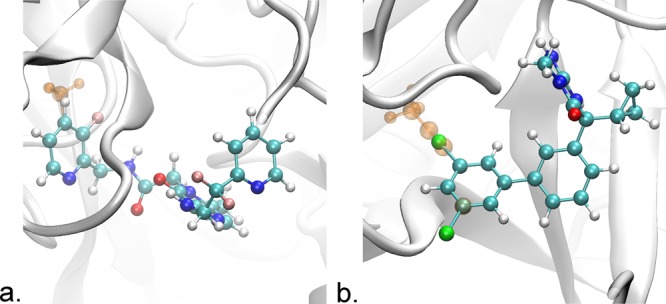
Figure 2.

Structures of the systems studied in this work. (a) Thrombin (gray ribbon) bound to the ligands CDA and CDB. The ligands differ in that CDB has a methyl group, represented here in orange, attached to the P1 pyridine ring. This ring has two possible conformations, one where the methyl group points In toward the protein as shown here and the other where the ring has flipped out. (b) BACE1 (gray ribbon) bound to the ligands 17a and 24. Ligand 17a has a 2,5-dichlorophenyl group, while ligand 24 has a 5-(Prop-1-yn-1-yl)pyridin-3-yl group. The atoms unique to ligand 24 are shown as transparent orange. This ring has two possible conformations, either In as shown or Out where it is flipped 180°. Tachyon28 in visual molecular dynamics29 was used for rendering.
