
Fig. 5.
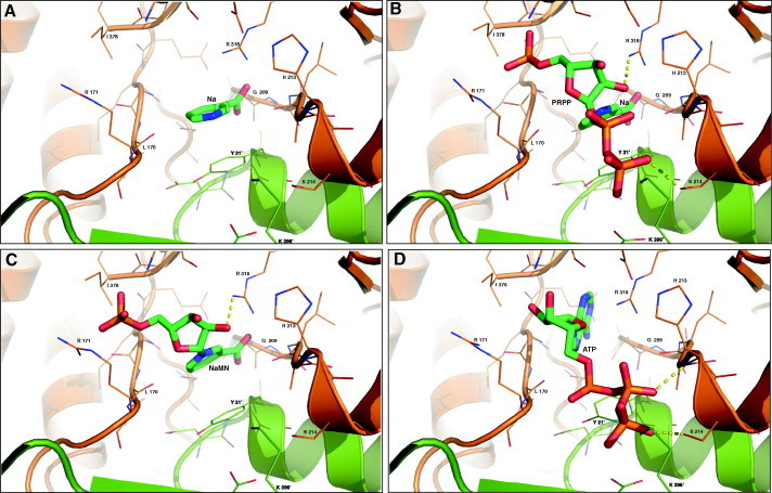
Molecular docking of the hNaPRTase active site dimeric interface in complex with different ligands. Side chains of residues participating in the binding ligands ((A) Na; (B) NaMN; (C) Na and PRPP; (D) ATP) are represented as thin sticks and their identity is indicated, whereas ligands are depicted as green sticks. Hydrogen bonds are shown as yellow dotted lines.