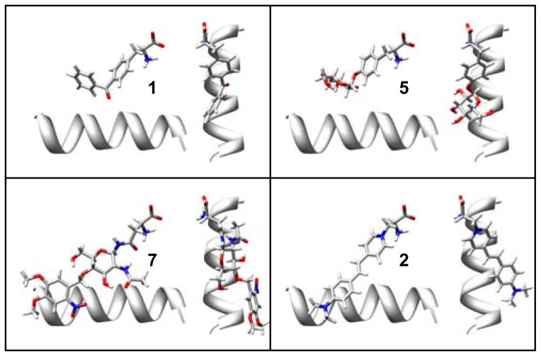
Figure 6.
Predicted ligand binding geometries of the best designs for 5, 7, and 2 compared to the crystallographic conformation of 1. The helix (res:150–163) is shown for reference. The binding geometry of 5 is very similar to 1. The 2-Nitro-4,5-DimethoxyPh protecting group of 7 can lay along the helix in an unstrained conformation. However, the requirement that 2 bound in a planar conformation was a significant complication that lead to less favorable designs.

