
Figure 4. Effect of accelerating flux through PDHC-NNT circuit on mitochondrial proton conductance.
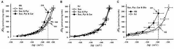
(A-C) Mitochondria were isolated from gastrocnemius and quadriceps muscle from C57BL/6N or C57BL/6J mice. (A & B) Graphs show rates of O2 consumption as a function of membrane potential (ΔΨm) in mitochondria isolated from C57BL/6N (+NNT; E) and C57BL/6J (-NNT; F) mice during respiration with minimal (succinate and succinate + pyruvate) and maximal (succinate + pyruvate + carnitine) flux through PDHC. Note the differences in JO2 (numbers with arrows) at a given ΔΨm (dotted line) indicating differences in proton conductance. (C) Direct comparison of proton conductance in isolated mitochondria from C57BL/6N and C57BL/6J mice under substrate conditions inducing maximal H2O2 production from PDHC and complex I (succinate + pyruvate + carnitine + glutamate). Note greater proton conductance in C57BL/6N mice at highest common ΔΨm.