
Figure 6.
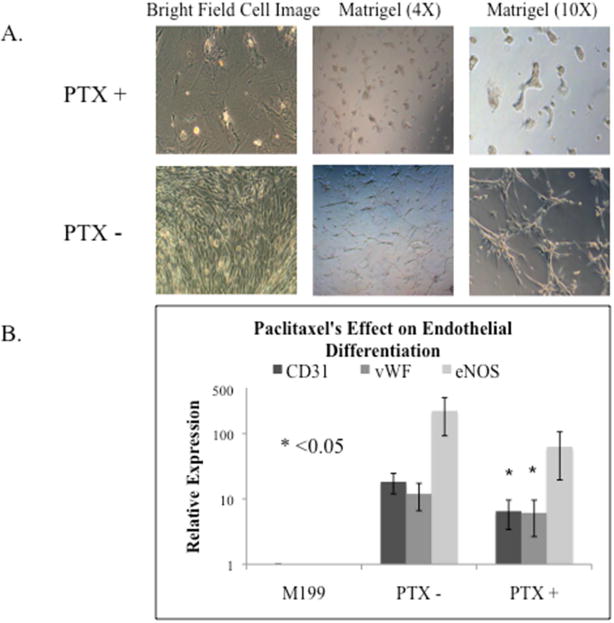
The effect of Paclitaxel (PTX) on hASC endothelial differentiation. A. Bright field cell images of control hASCs and Paclitaxel treated hASCs in 6-well plates and Matrigel coated 24-well plates. Light microscopy showed Paclitaxel treatment induced cell morphology changes and less tube formation. B. The expression of endothelial differentiation markers CD31, vWF, and eNOS, measured by Real-time PCR. Two of the three endothelial markers, CD31 and vWF, revealed significantly reduced expression in Paclitaxel treated hASCs (p=0.016, p=0.031, p=0.500).