Figure 6.
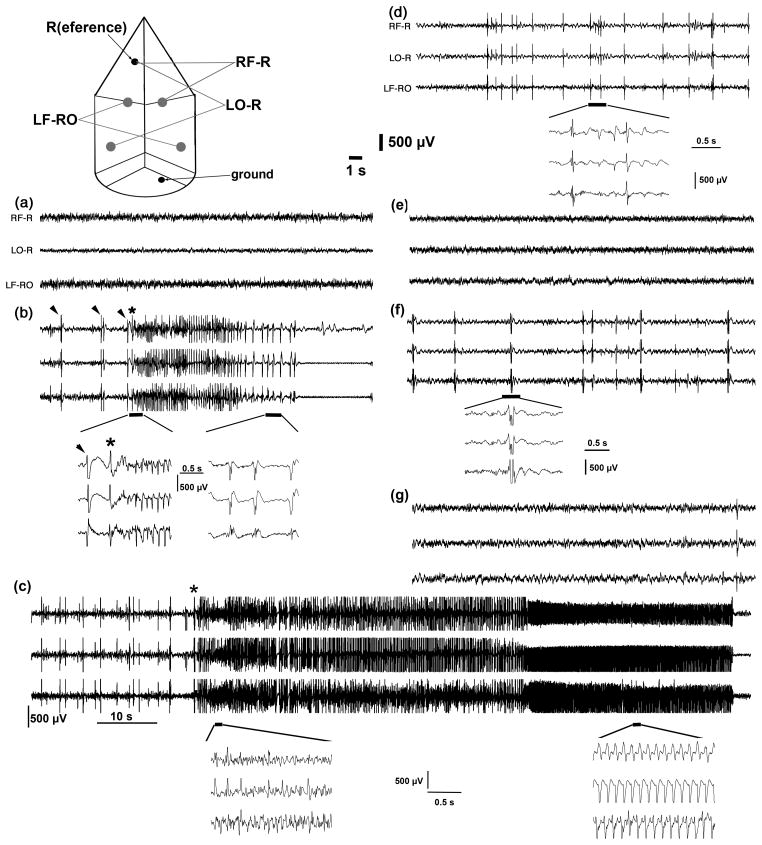
EEG recordings from mice treated by simultaneous (a–d) or sequential (e–g) DZP:MK-801 3:1 regimens after TMDT poisoning Top - scheme of cortical electrodes and montages. There were two active electrodes over the frontal (sensorimotor) cortex and two electrodes over the occipital (visual) cortex. Reference electrode was represented by a screw in the nasal bone, a screw above the cerebellum served for the purpose of grounding. RF-R = right frontal vs. reference; LO-R = left occipital vs. reference. LF-RO = left frontal vs. right occipital. Time mark 1 s and calibration 500 μV for all traces except if noted otherwise.
(a) Baseline (pre-TMDT) EEG recording in a mouse later injected with TMDT (0.4 mg/kg i.p.) and after the first clonic seizure with simultaneous diazepam (1.2 mg/kg ip) + dizocilpine maleate (0.4 mg/kg i.p.; DZP+MK-801).
(b) Synchronous EEG discharges of spike-and-wave character (marked by arrowheads and associated behaviorally with whole body twitches) and an EEG seizure (asterisk at onset; poly-spike, later spike-and-wave) associated with clonic behavioral seizure after TMDT in the same mouse. This clonic seizure was the first distinct feature of TMDT poisoning and treatment (DZP + MK-801) has been initiated immediately afterwards (within 10 s). First inset shows onset of clonic seizure with longer time base. Please note the first spike followed by a slow wave (marked by an arrowhead) followed after the second spike-wave complex (*) by a burst of spikes (polyspikes). Second inset shows polyspike-and-wave EEG pattern at the end of this clonic seizure.
(c) Prolonged EEG discharges corresponding to a clonic seizure (onset marked with an asterisk; duration 90 s) in the same mouse now about 2 hours after the combined (simultaneous) DZP and MK-801 treatment. Clonic seizures longer than 30 s regularly occurred in the mice treated simultaneously. Time mark 10 s, calibration 500 μV. First inset (with own time mark and calibration) shows a period from the onset of this clonic seizure characterized by frequent spikes, although not clearly developed in all traces. Second inset demonstrates typical rhythmical spike-wave complexes from the terminal part of this prolonged seizure.
(d) Profound attenuation of background EEG activity with interictal discharges in the same mouse about 15 min prior to demise at 7 hours after TDMT poisoning. Inset shows several of these discharges characterized by very short polyspikes, sharp wave-slow wave complexes, sharp waves, and spike-wave complexes.
(e) Baseline (pre-TMDT) EEG recording in a mouse later injected with TMDT (0.4 mg/kg i.p.) and DZP followed in 10 min with MK-801 (sequential treatment). Baseline EEG activity was analogous in all mice, please compare with (a).
(f) Frequent interictal EEG discharges accompanied by whole body twitches represented a hallmark of TMDT toxicity in the mice treated with sequential DZP (1.2 mg/kg i.p.) and MK-801 (0.4 mg/kg). Inset reveals the character of interictal discharges as polyspike-wave complexes.
(g) Almost complete recovery of EEG activity (cf. to (e)) in the mouse at 24 hours after TMDT injection and sequential treatment with DZP and MK-801. Please note a single interictal discharge at the end of the recording.

