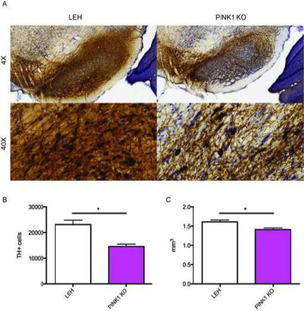
Fig. 1.
Analysis of nigral dopaminergic loss in the PINK1 KO animals. Brain tissue was isolated, sectioned, and stained for tyrosine hydroxylase (TH) (A). The number of TH-positive neurons (B) and volume (C) were quantified for the substantia nigra pars compacta. *: p≤0.05 on a t-test. n=6 animals for both groups.

