Fig 1.

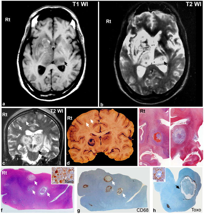
a,b: Axial T1WI and T2WI taken at the level of basal ganglia show multiple lesions involving the bilateral thalami, left caudate, right lentiform nucleus and left crus cerebri (arrow, b). Lesions in the thalamus appear hypointense on T1WI (a) with corresponding concentrically hypo and hyper intense zones on T2WI (“concentric target” sign) (b) with perilesional edema. Small punctate hyper intensities seen within the lesion on T1WI and ring like hyperintense left caudate lesion (a) suggests the presence of paramagnetic material/blood.
c: Coronal T2WI at the level of bilateral thalami and crus cerebri shows multiple lesions involving the bilateral thalami, and crus cerebri with perilesional edema. The thalamic lesions revealed “concentric target like” appearance with multiple layers of alternate concentric hypo- and hyper intensities with central hypointensity. Similar lesion also seen in left crus cerebri (black arrow). Smaller hyperintense lesions are noted in the bilateral superior frontal and cingulate gyrus at grey white junction (white arrows). Some of these have central/eccentric hypointensities.
d,e: Gross coronal slice of brain at same level as MRI revealed well circumscribed lesions in bilateral thalamus (d). The lesion on the right side had concentric zones of hemorrhage separated by pale necrotic zones, while the left was necrotizing with a thin outer and inner hyperaemic rim. Multiple smaller abscess like lesions (0.5 cms across) are seen along the grey white junction of the right superior frontal gyrus (d, arrows).
Large format whole mount histologic preparation from bilateral basal ganglia (e) taken at same level as lesions on MRI for comparison reveal large hemorrhagic and necrotizing lesions.
f-h: The lesions in right frontal cortex are organising abscesses with central zones of coagulative necrosis (f) and surrounding histiocytes (g). Tachyzoites and bradyzoites of T gondii at the periphery of lesion is seen by immunohistochemistry (f, inset). Note accumulation of tachzoites of T gondii seen rimming the lesion in crus cerebri on immunohistochemsitry (h). Inset reveals numerous ruptured tachyzoites surrounding inflamed vessels.
[e: Whole mount preparation, Masson trichromex8, f: HEx10, f, inset: Toxoplasma immunostain xObj.40, g: Cd68 immunostainx10, h: Toxoplasma immunostainx10, h,inset: Toxoplasma immunosatinxObj.20]
