Figure 4.
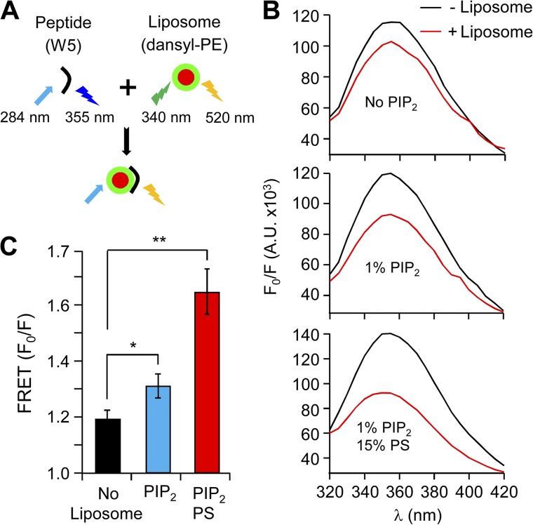
Binding of the N-terminal peptide of β2e to liposomes is strengthened in the presence of anionic poly-PIs. (A) Schematic diagram of FRET analysis. Binding was examined using FRET between peptide containing Trp (W) (donor) and liposomes labeled with dansyl-PE (acceptor). Fluorescence emitted from Trp is quenched by dansyl-PE, suggesting peptide binding to liposomes. The initial spectrum of Trp was determined in the absence of liposomes (F0), and the subsequent spectrum was recorded after liposome addition (F). (B) FRET signals in the absence (black trace) or presence (red traces) of liposomes with no-liposome, 1% PIP2, or both 1% PIP2 and 15% PS. FRET is presented as F0/F at 355 nm. A.U., absorbance units. (C) Summary of FRET changes with different lipid compositions on the liposomes. For no-liposome, 1% PIP2, and both 1% PIP2 and 15% PS, n = 3; *, P < 0.05; **, P < 0.01, compared with no liposome. Data are mean ± SEM.

