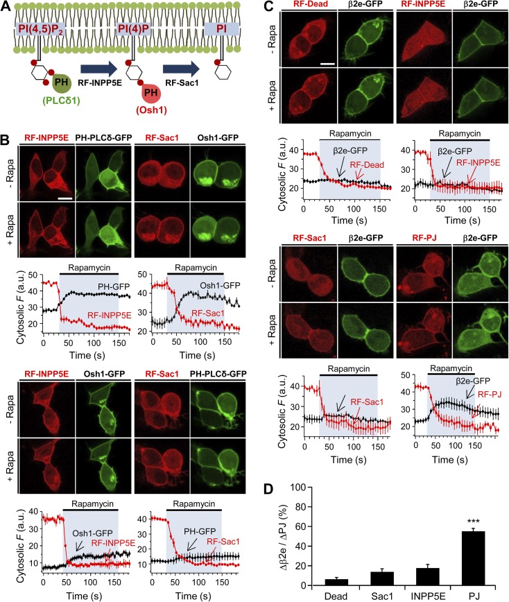
Figure 5.
Depletion of PI(4)P and PIP2 induces the dissociation of β2e subunit from the plasma membrane. (A) Schematic diagram of a depletion of PI(4)P or PIP2 by 4- or 5-phosphatases. (B) Confocal images and full time courses of cytosolic fluorescence change in cells transfected with LDR, PH-PLCδ-GFP, Osh-1-GFP, RF-INPP5E, and RF-Sac. Translocation of RF-INPP5E or RF-Sac to the plasma membrane by rapamycin depletes PH-PLCδ-GFP (PIP2 probe) or Osh1-GFP (PI(4)P probe), respectively. For INPP5E, n = 3; for Sac, n = 3. Bottom images show that plasma membrane translocation of RF-INPP5E or RF-Sac by rapamycin has no effect on depletion of Osh1-GFP or PH-PLCδ-GFP probes, respectively. For INPP5E, n = 3; for Sac, n = 3. Time courses were taken every 5 s by confocal microscope. Bar, 10 µm. (C) Confocal images and full time course of cytosolic fluorescence change in cells transfected with LDR, β2e-GFP, and RF-Dead (RFP-FKBP-dead form of PJ), RF-INPP5E, RF-Sac, and RF-PJ. Confocal images show the subcellular distribution of β2e-GFP and translocatable RF-phosphatases before and after rapamycin (1 µM) application for 2 min. For Dead, n = 4; for INPP5E, n = 3; for Sac, n = 3; for PJ, n = 4. Bar, 10 µm. (D) Change in cytosolic intensity of β2e subunit (Δβ2e) to cytosolic intensity of each translocatable enzyme (ΔPJs) before and after rapamycin application and expressed as percentage with Dead (n = 5), Sac (n = 5), INPP5E (n = 5), and PJ (n = 6). ***, P < 0.001, compared with Dead. Data are mean ± SEM.

