Figure 7.
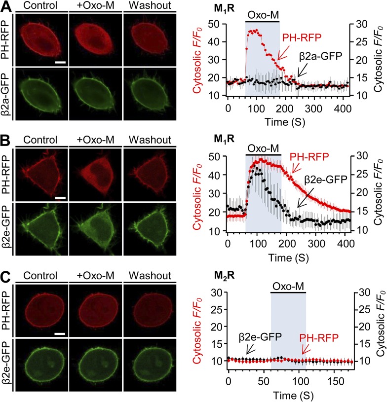
Activation of M1R induces reversible translocation of the β2e subunit. Confocal images and full time courses of PH-PLCδ-RFP (PH-RFP) and β2a-GFP (A) or PH-RFP and β2e-GFP (B) before, during, and after Oxo-M (10 µM) application in M1R-expressing cells. Time courses were taken every 5 s by confocal microscope. (C) Subcellular distributions and a full time course of PH-RFP and β2e-GFP in M2R-expressing cells. For analysis of time course, n = 4–5. Scale bars, 10 µm.

