Figure 1.
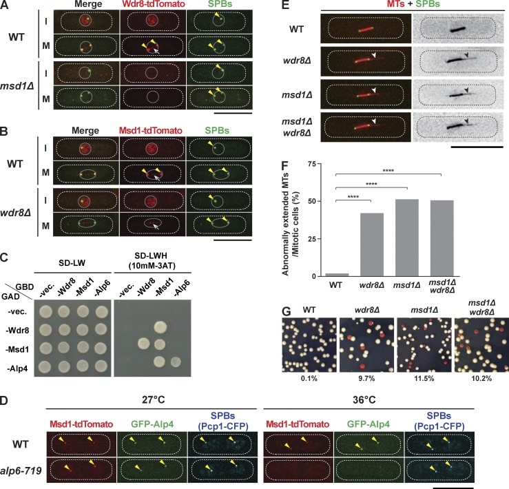
Wdr8 is required for anchoring spindle microtubules to the SPB. (A and B) Mitosis-specific SPB localization of Msd1 and Wdr8 is interdependent. Representative images of wild-type (WT) and msd1Δ mutant cells containing Wdr8-tdTomato and GFP-Alp4 (SPB; A) and of wild-type and wdr8Δ mutant cells containing Msd1-tdTomato and GFP-Alp4 (B) are shown. Localization of individual proteins during interphase (I; top) or mitosis (M; bottom) is shown in each row. Cells were grown in rich media at 27°C. The positions of SPBs and spindle microtubules are indicated with arrowheads and arrows, respectively. The peripheries of the cell and the nucleus are outlined (dotted and continuous lines, respectively). (C) Wdr8 interacts with Msd1. Yeast two-hybrid assay was performed with the indicated plasmids containing Gal4 activation domain (GAD) and Gal4 DNA-binding domain (GBD). Interaction was assessed according to growth on minimal complete synthetic defined plates lacking leucine and tryptophan (left, −LW) or leucine, tryptophan, and histidine but containing 3AT (right, −LWH, 10 mM 3AT). vec., vector. (D) Alp4 and Msd1 are delocalized from the SPB in the alp6-719 mutant. Wild-type and alp6-719 mutant cells containing Msd1-tdTomato, GFP-Alp4, and Pcp1-CFP, an SPB marker (Flory et al., 2002; Fong et al., 2010), were grown at 27°C (left), shifted to 36°C, and kept at that temperature for 2 h (right). Representative images of each strain are shown. The positions of SPBs are indicated with arrowheads. (E) wdr8Δ mutants display protruding spindle microtubules. Morphology of mitotic spindle microtubules in wild-type, wdr8Δ, msd1Δ, and msd1Δwdr8Δ mutant cells containing mCherry-Atb2 (microtubules [MTs]) and GFP-Alp4 (SPBs) are shown. Cells were grown in rich media at 27°C. The protrusion of spindle microtubules is indicated with arrowheads. (F) Quantification. The percentage of mitotic cells displaying abnormally extended microtubules is quantified. All p-values were obtained by performing the two-tailed χ2 test (≥40 cells). We followed this key for asterisk placeholders for p-values in the figures: ****, P < 0.0001. (G) wdr8Δ mutants show a minichromosome loss phenotype. Indicated strains carrying the minichromosome Ch16 (Niwa et al., 1989) were grown on rich yeast extract plates (lacking adenine) and incubated at 30°C for 4 d. Cells that had lost the minichromosome formed red or red-sectored colonies. The percentages of red-sectored colonies are shown at the bottom (n ≥ 1,000). Bars, 10 µm.

