Figure 5.
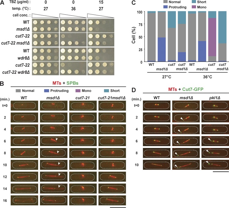
Mutual suppression of defective phenotypes in double mutants between cut7 and msd1 or wdr8. (A) Suppression of temperature and TBZ sensitivity. Serial dilution spot tests were performed by using the indicated strains on rich agar media in the presence or absence of thiabendazole (TBZ), and the cells were incubated at the indicated temperatures for 3 d. cell conc., cell concentration. (B) Time-lapse images showing mitotic progression and spindle microtubule morphology. Live imaging of individual strains that contained mCherry-Atb2 (microtubules [MTs]) and GFP-Alp4 (SPBs) was performed at 36°C. Three distinct characteristic phenotypes were identified (protruding spindles, msd1Δ; monopolar spindles, cut7-21; short spindles, cut7-21msd1Δ). Representative images from wild-type (Video 1), msd1Δ (Video 2), cut7-21 (Video 3), and cut7-21msd1Δ (Video 4) cells are shown. Arrowheads show protruding spindle microtubules. (C) Quantification of phenotypes in each mutant. Cells that spent ≥10 min with short spindles (see the cut7-21msd1Δ cell in B) were classified as short. At least 20 mitotic cells of each strain were observed. (D) Abnormal localization of Cut7 in msd1 or pkl1 deletion mutants. Time-lapse imaging of Cut7-GFP and mCherry-Atb2 (microtubules) in wild-type, msd1Δ, and pkl1Δ cells is shown. Cut7-GFP signals accumulating close to the tips of the protruding spindle microtubules are marked by arrowheads in the msd1Δ or pkl1Δ mutant cell. The peripheries of the cell are outlined in the images with dotted lines. WT, wild type. Bars, 10 µm.

