Figure 6.
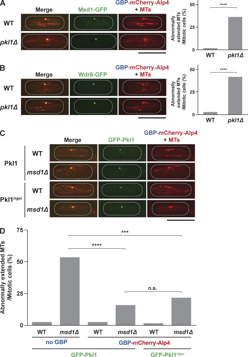
Tethering of Pkl1, either wild type or the rigor mutant, to the SBP is largely sufficient for spindle anchoring. (A) Tethering Msd1 to the SPB is not sufficient for spindle anchoring in the absence of Pkl1. Representative interphase images of Msd1-GFP, GBP-mCherry-Alp4 signals, and mCherry-Atb2 (microtubules [MTs]) in the wild-type (top) and pkl1Δ mutant (bottom) cells are shown. (left) Note that Msd1-GFP is colocalized with the SPB/Alp4 during interphase. Quantification of spindle-anchoring defects is shown on the right. P-value was obtained from the two-tailed χ2 test (≥50 cells; ****, P < 0.0001). (B) Tethering Wdr8 to the SPB is not sufficient for spindle anchoring in the absence of Pkl1. Representative interphase images as in A are shown except that the localization of Wdr8-GFP is displayed. P-value was obtained from the two-tailed χ2 test (≥50 cells; ****, P < 0.0001). (C) Tethering of Pkl1 to the SPB in wild type or msd1Δ. GFP-Pkl1, either wild type (top two rows) or the rigor mutant (bottom two rows), was tethered to the SPB by using GBP-mCherry-Alp4. Representative interphase images as in A are shown except that the localization of GFP-Pkl1 is displayed. (D) Tethering Pkl1, either wild type or rigor, to the SPB alone significantly rescues spindle-anchoring defects in msd1Δ. All p-values were obtained from the two-tailed χ2 test (≥50 cells; ***, P < 0.001; ****, P < 0.0001; n.s., not significant). The peripheries of the cell are outlined in the images with dotted lines. WT, wild type. Bars, 10 µm.

