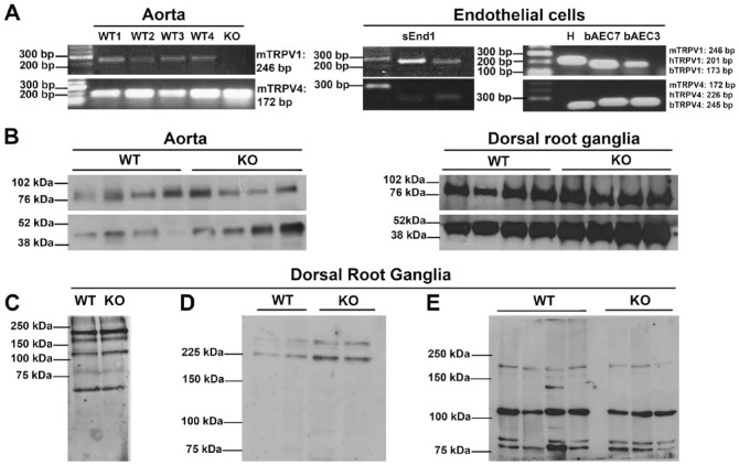
Figure 1.
(A) Murine [m], human [h] and bovine [b] TRPV1 and TRPV4 mRNA in aortic lysates from four TRPV1 wild-type (WT1–4) mice and one TRPV1 knock out (KO) mouse (left panel), and in mouse skin endothelioma cells (sEnd1), human umbilical vein endothelial cells (H), and bovine aortic endothelial cells at passage 7 (bAEC7) and passage 3 (bAEC 3) (right panel). (B) Representative immunoblots of TRPV1 protein expression in murine aortic and dorsal root ganglia lysates from TRPV1 WT and KO mice, probed with ACC-030 anti-TRPV1 antibody (Alomone Labs, Jerusalem, Israel; predicted molecular weight, 95 kDa). β-actin expression was used as a loading control (predicted molecular weight, 42 kDa). (C–E) Representative immunoblots of dorsal root ganglia lysates from TRPV1 WT and KO mice using a number of different anti-TRPV1 antibodies: (C) ab4579, Abcam (Cambridge, UK); (D) ACC-030 Batch 2, Alomone Labs; (E) V2764, Sigma-Aldrich (St Louis, MO).

