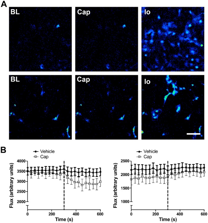
Figure 2.
(A) Capsaicin-induced calcium fluorescence in murine pulmonary endothelial cells (upper panel) and murine aortic smooth muscle cells (lower panel). Representative images were captured at baseline (BL), and after stimulation with 1 µM capsaicin (Cap), and 1 µM ionomycin (Io). No increase in intracellular Ca2+ was observed in either endothelial or smooth muscle cells in response to 1 µM capsaicin. Scale, 40 µM. (B–C) Blood flow responses in first-order mesenteric vessels treated with capsaicin and vehicle (2% DMSO in saline) in healthy and endotoxaemic (LPS; 12.5 mg/kg, i.v., 24 hr) wild type (WT) mice, respectively. Baseline mesenteric blood flow was recorded for 5 min; capsaicin (Cap; 10 µM) or vehicle (2% DMSO in saline) was then administered as an aerosolized spray, denoted by the dotted line, and blood flow was recorded for a further 5 min. In naïve mice, capsaicin caused a decrease in blood flow, indicative of vasoconstriction; in LPS-treated mice, however, capsaicin increased blood flow. Data are presented as mean ± SEM (n=6–14).

