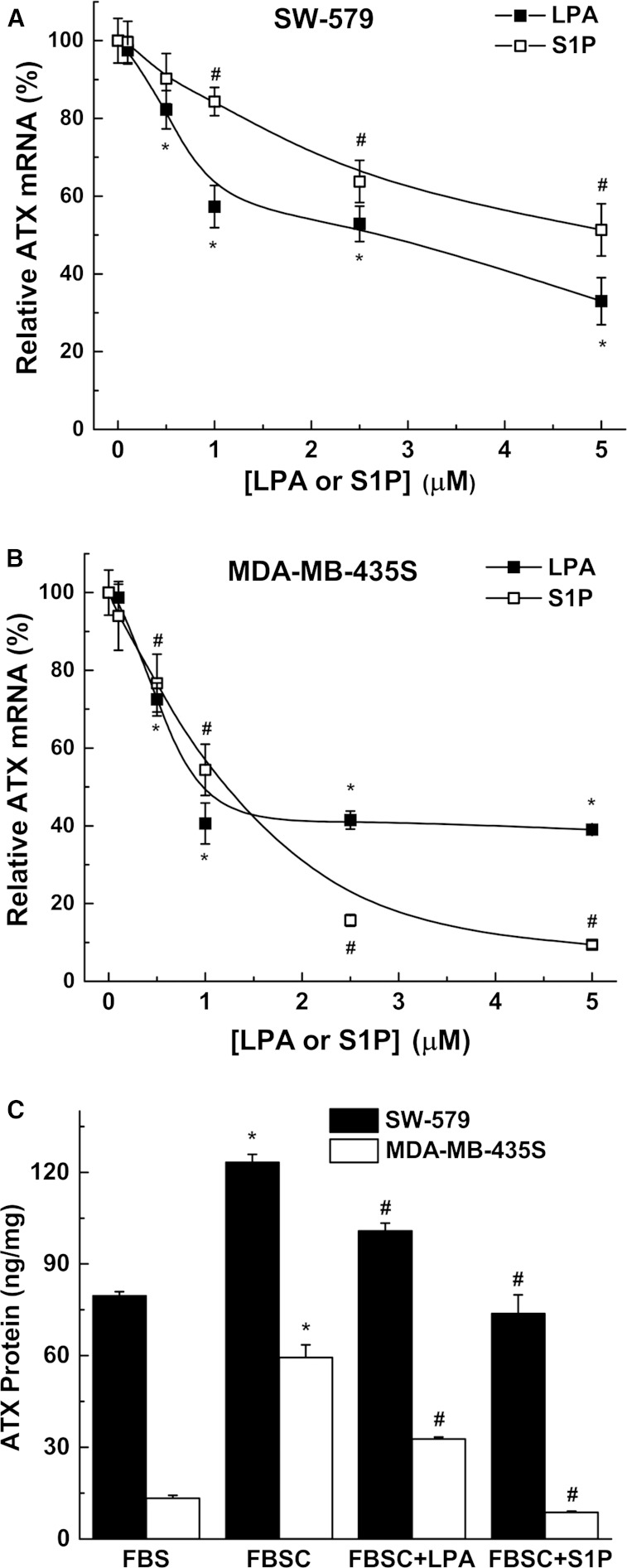
Fig. 3.
LPA and S1P decrease ATX mRNA expression and secretion of ATX. SW-579 (A) and MDA-MB-435S (B) cells were incubated for 24 h in 10% FBSC medium with increasing concentrations of either LPA or S1P. Significant reduction (P < 0.05) in ATX mRNA expression with LPA (*) and with S1P (#) compared with no treatment. C: SW-579 and MDA-MB-435S cells were incubated for 48 h in medium containing FBS or FBSC with or without 5 μM LPA or S1P. Secreted ATX protein was quantified by ELISA and normalized to both volume of conditioned medium and cell lysate protein content. ATX protein from basal medium was subtracted from the total ATX protein measured after incubation. Results are mean ± SEM from three independent experiments. *A significant increase (P < 0.05) in ATX protein concentration in FBSC medium compared with FBS medium. #A significant decrease (P < 0.05) in ATX protein following LPA or S1P treatment in FBSC compared with FBSC alone.

