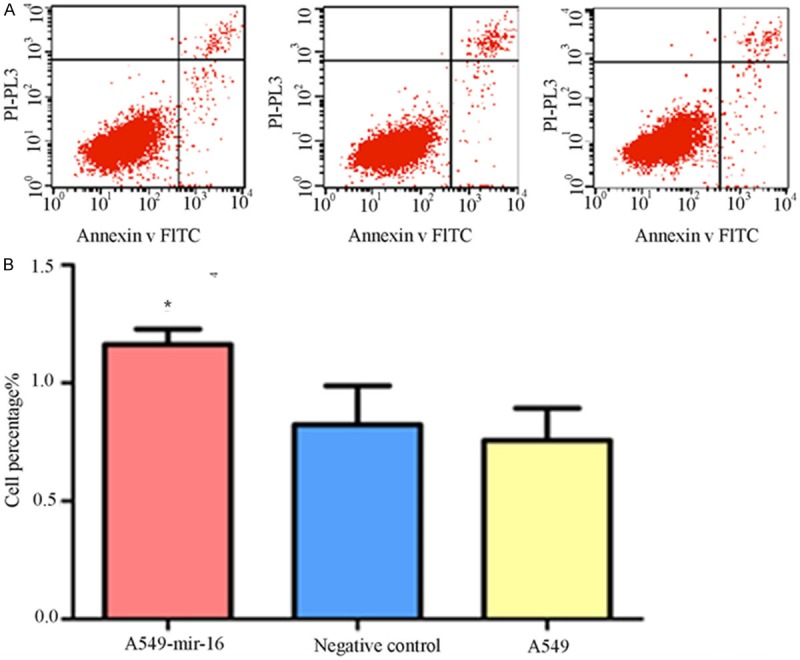
Figure 3.

Effect of mir-16 on cell apoptosis. A. Flow cytometry pictures of cell apoptosis (lower right quadrant, one of the three separate results was showed); B. Percentage of three groups in early stage apoptosis. Flow cytometry showed that the percentage of mir-16 transfected A549 cells in early stage apoptosis is higher than that of other two groups (values represent means from three separate experiments, *P<0.05).
