
Figure 2.
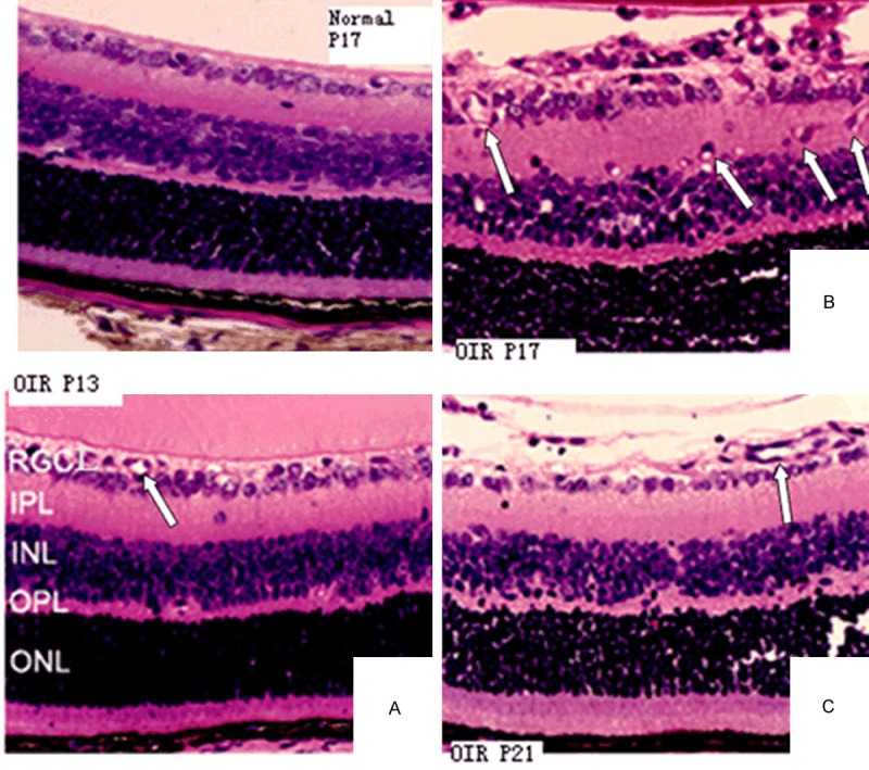
H&E staining of the retinas from the control group (P17) shows a limited number of vascular endothelial cells and the lumen-sample structure in retinal internal limiting membrane (400×). H&E staining of the retinas from the OIR group on P13, P17 and P21 (400×). A. On P13, the internal limiting membrane was integrated and blood vessels were dilated. B. On P17, the internal limiting membrane was not integrated. Large amounts of visible vascular endothelial cell nuclei, alone or clustered, broke through the retinal inner limiting membrane. C. On P21, the internal limiting membrane became smoother. The number of vascular endothelial cells breaking through the internal limiting membrane was significantly reduced. Arrows indicate newly formed blood vessels.