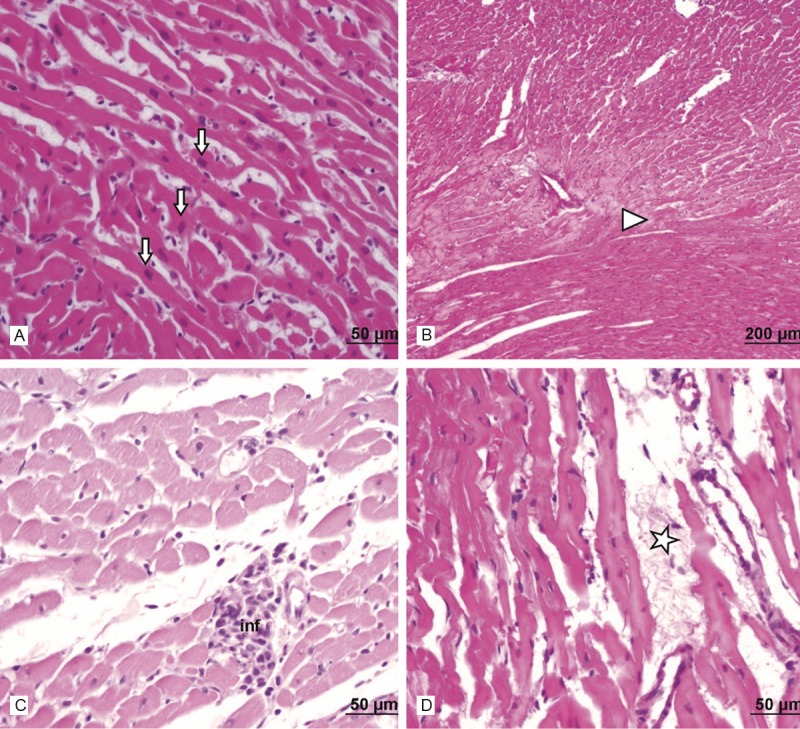
Figure 4.

Assessment of heart light microscopy analysis in 30 mg- kg mg- kg azithromycin-administered rats. In the heart tissues of azithromycin-administered rats, light microscopic examination showed necrosis (►) and hypertrophy (↓) of cardiac muscle cells, infiltration of inflammatory cells (inf) and interstitial edema (*). A. Haematoxylin and eosin stain, scale bar-50 μm; B. Haematoxylin and eosin stain, scale bar-200 μm; C. Haematoxylin and eosin stain, scale bar-50 μm; D. Haematoxylin and eosin stain, scale bar-50 μm.
