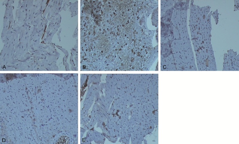
Figure 3.

Expression of PKD1 in myocardial tissues. Immunohistochemical staining was performed to investigate the expression of PKD1 in cardiomyocytes. Cells stained brown were PKD1-positive.The expression of PKD1 in cardiomyocytes of the sham-operated group (A), the control group (B), the Astragalus group (C), the Salvia group (D), and the compatibility of Astragalus and Salvia group (E) were visualized by an optical microscope (× 100).
