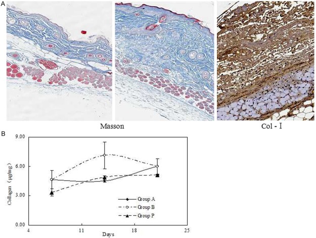
Figure 1.
Collagen content and the effect of APS treatment at different time. A. Collagen in the dermis was observed Masson’s trichrome stains and immunohistochemistry of type I collagen in the BLM-treated mice. B. APS was administered to mice intravenously after initiating BLM treatment and contents of hydroxyproline were detected to determine collagen content.

