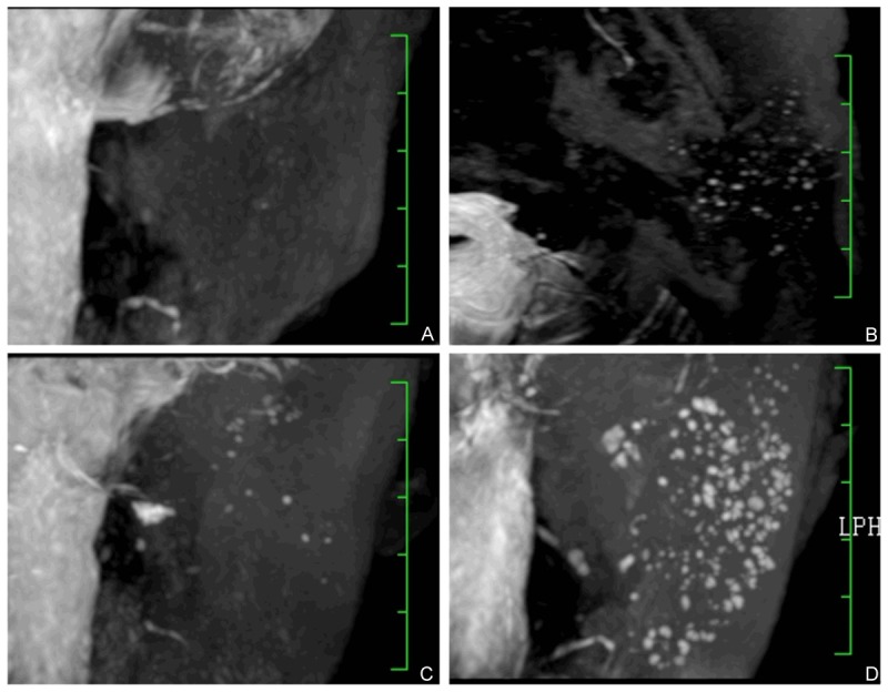
Figure 2.

3D-T2-DRIVE MR sialography grading. Images showed grade 1 to 4 duct dilation. A: Grade 1, punctate areas of high-signal intensity, 1 mm or less in diameter. B: Grade 2, globular spherical areas of high signal intensity, 1 to 2 mm in diameter. C: Grade 3, cavity-like areas of high intensity coalesce and enlarge further, more than 2 mm in diameter. D: Grade 4, marked dilation of the main duct with an irregular diameter, as well as irregular branching.
