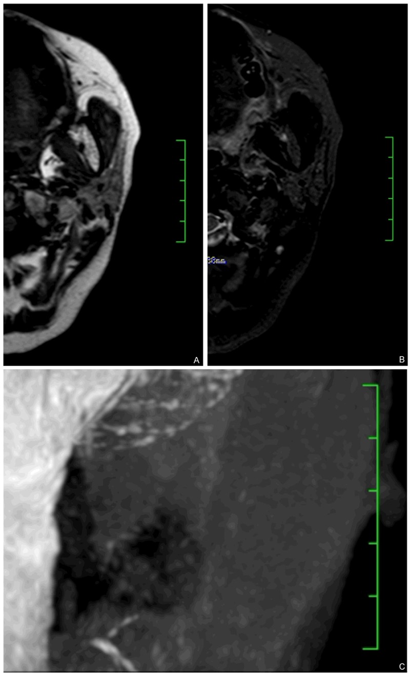
Figure 3.

Combination of MRI imaging and 3D-T2- DRIVE MR sialography identified unparalleled fat deposition and ducts dilation. (A and B) showed axial T1WI and STIR showing diffusive fat signal, more than 50% of whole parotid glands, barely see any normal gland structure. The results indicated this patient is fat signal grade 4. However, in the same patient, MR sialography image (C) showed moderate grade 2 peripheral duct dilation.
