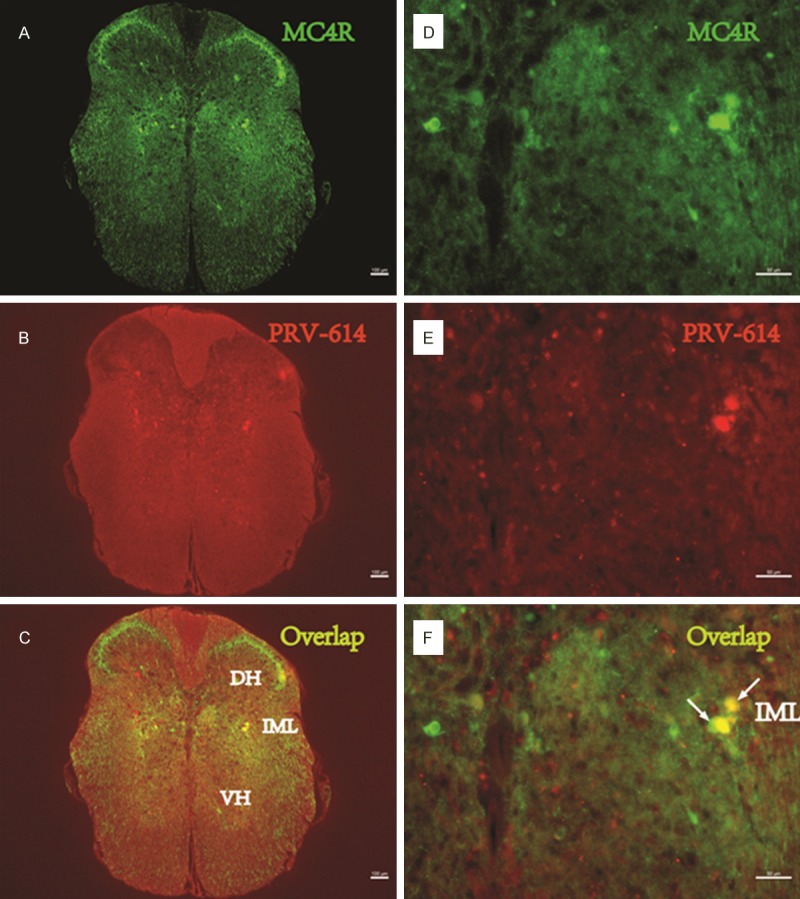
Figure 1.

PRV-614/MC4R-GFP double-labeled neurons in the spinal cord. Images (A-C) were taken from an animal after injections of PRV-614. (A) Showed MC4R-GFP positive neurons in the spinal cord; (B) Showed neurons infected with PRV-614, which send transsynaptic projections to the stomach; (C) Showed overlaid images of (A) plus (B). Images (D-F) amplified views of (A-C), respectively. Arrows (white) indicate double-labeled neurons. IML, the intermediolateral cell column; DH, Dorsal horn; VH, ventral horn. Scale bars, 50 μm.
