Figure 1.

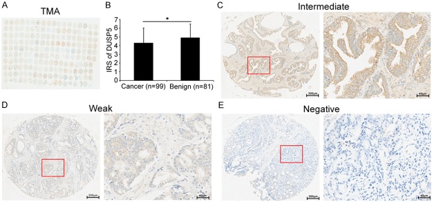
Immunohistochemical staining for DUSP5 in PCa and adjacent non-cancerous prostate tissues in our TMA samples. (A) A full view of the immunohistochemistry staining for DUSP5 in our TMA cohort. (B) The immunoreactivity scores (IRS) of DUSP5 in prostate cancer were lower than that in adjacent benign prostate tissues (IRS: PCa = 4.29 ± 1.72 versus Benign = 4.89 ± 1.58, P = 0.04). Data were presented as Mean ± SEM. *P < 0.05. (C-E) The immunohistochemistry staining indicated that DUSP5 immunostainings mainly occurred in the cytoplasm and cellular membrane of PCa and benign glandular epithelium cells. The intensity of DUSP5 immunostainings was intermediate (C), weak (D) and negative (E), with the exception of strong (Left panel: magnification × 40; right panel: magnification × 200).
