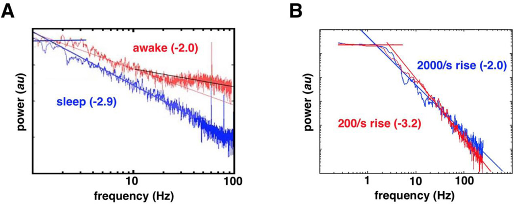
Fig. 2. Evidence for spectral slope changes.
(A) During sleep the human subdural ECoG PSD has a steep negative slope that flattens (whitens) during wakefulness. (B) Simulations of human ECoG PSD suggest that PSD slope changes arise as a function of the dendritic response to an input. As the strength of the input increases, more neurons fire simultaneously, forcing a greater local population to become refractory within a shorter time window, increasing the rise time. Thus, for weaker inputs with greater rise times the slope of the PSD is flatter (blue), whereas for stronger inputs with smaller rise times the slope of the PSD is steeper (red). Thus, within our framework, if the dendritic response is driven by ephaptic coupling, the stronger the coupling, the steeper the slope; with weaker ephaptic coupling, spike times are less temporally correlated and the slope is flatter. (From Freeman & Zhai, 2008)(68)

