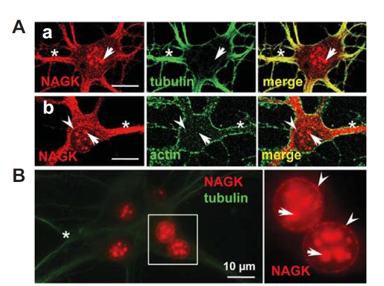
Fig. 1.

Nuclear expression of NAGK in hippocampal neurons. Cultured rat hippocampal neurons (DIV 21) were double-labeled with indicated antibodies. (A) ICC. Double-labeling with anti-NAGK/-tubulin (a) or with anti-NAGK/actin (b) revealed the expression profile of NAGK was similar to that of tubulin in dendroplasm (asterisks). NAGK clusters in the central area of soma (arrows) and a circular arrangement (arrowheads) were marked. (B) INC. Double-staining ‘naked’ nuclei with anti-NAGK/-tubulin antibodies revealed patch-like NAGK-IR signals in nucleoplasm (arrows) with a circular distribution (arrowheads). The boxed area is enlarged in the inset on the right. Scale bar; 10 μm.
