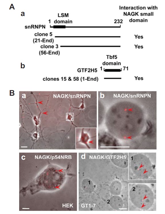
Fig. 5.

Interaction of NAGK with nuclear proteins. (A) Yeast two hybrid assays using the NAGK small domain as bait identified snRNPN and GTF2H5. The NAGK-interacting domains of snRNPN (a) and GTF2H5 (b) of two different clones for each protein are shown. The positions of like-Sm (LSM) and TTDA subunit of TFIIH basal transcription factor complex (Tbf5) protein domains of each protein were marked on the bars with amino acid numbers. (B) PLA showing interactions of NAGK with nuclear proteins. PLA was performed in rat hippocampal neurons (DIV 11). A low power image shows distribution of NAGK/snRNPN PLA dots (arrowheads) in soma (inset) and dendrites (a). Immunonucleochemistry followed by PLA shows the interaction between NAGK and snRNPN (arrows) in the ‘naked’ neuronal nucleoplasm (b). Scale bars: 5 and 20 μm in a and b, respectively. PLA conducted in HEK293T cells showed interaction between NAGK and paraspeckle protein (p54NRB) in nucleus (c, arrowheads). Scale bar, 5 μm. PLA done in hypothalamic GT1-7 cells showed NAGK/TTDA (GTF2H5) PLA dots in nucleoplasm (arrowheads). Scale bar, 10 μm.
