
Fig. 1.
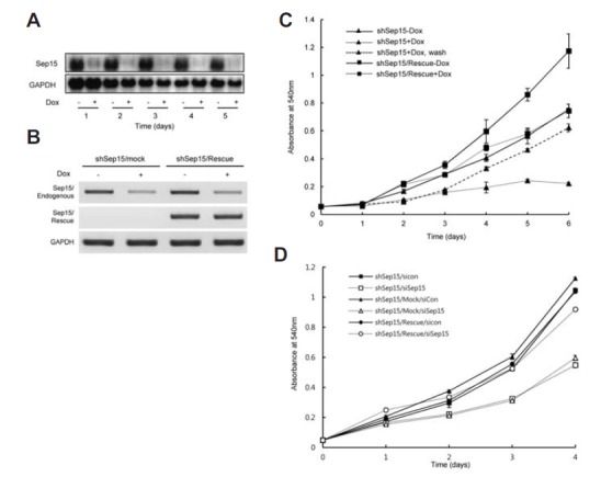
Sep15 deficiency leads to the inhibition of cell proliferation. (A) Efficiency of Sep15 knockdown after the induction of shRNA expression was measured by northern hybridization. GAPDH was used as an internal control. (B) Measuring the endogenous and rescue-specific Sep15 mRNAs by RT-PCR. cDNAs were amplified using specific primer sets as described in Materials and Methods. (C) Cell proliferation in shSep15-Dox cells (triangles connected with a solid line), shSep15+Dox (triangles connected with a dotted line), removal of Dox 3 days after induction of knockdown (triangles connected with a discontinuous line), shSep15/Res- cue-Dox cells (squares connected with a solid line), and shSep15/Res- cue+Dox cells (squares connected with a dotted line) were measured by a MTT assay. Data shown are representative of at least three independent experiments. (D) The effect of Sep15 knockdown using siRNA on cell proliferation. shSep15 cells were transfected with a solid line) or siSep15 (open squares with a dotted line), shSep15/Mock cells containing both shSep15 and rescue backbone vector (shSep15/Mock) with siCon (closed triangles with a solid line) or siSep15 (open triangles with a dotted line), and shSep15/Rescue cells with siCon (closed circles with a solid line) or siSep15 (open circles with a dotted line). Cell proliferation was measured by a MTT assay. Data shown are representative of at least three independent experiments and error bars stand for standard deviations.