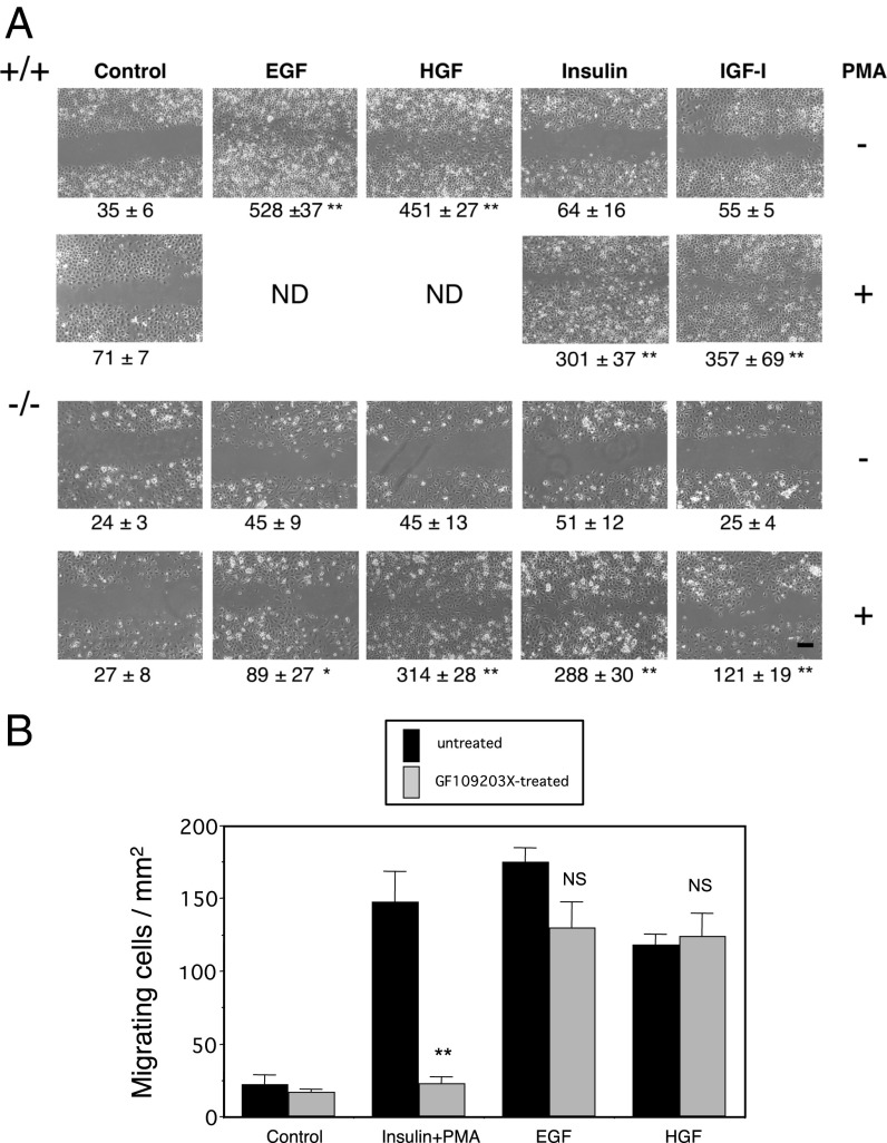
Fig. 4.
PMA promotes in vitro keratinocyte migration independent of Stat3. (A) Primary cultured keratinocytes were subjected to in vitro migration assays in the presence of the indicated growth factors. Every assay was performed after treating keratinocytes with mitomycin C to prevent their proliferation. Stat3+/+ keratinocytes showed strong migration in response to EGF or HGF (Top) as previously described (10) but not in response to insulin or IGF-I (Top). In contrast, Stat3−/− keratinocytes did not migrate in response to any growth factor tested (third row). PMA synergistically stimulated the migration of Stat3+/+ (second row) and Stat3−/− (Bottom) keratinocytes in the presence of insulin or IGF-I. In addition, Stat3−/− keratinocytes slightly but significantly migrated in response to PMA plus EGF (P < 0.05) and considerably to PMA plus HGF (Bottom). Note that treatment with PMA alone did not stimulate migration of Stat3+/+ or Stat3−/− keratinocytes (second and third rows). ND, not done. (Bar = 200 μm.) Quantitative evaluation of cell migration (see Materials and Methods) is shown below each panel. Significantly different from the control (*P < 0.05; **P <0.01) as determined according to Student's t test. (B) Although PMA plus insulin-induced migration of wild-type keratinocytes was completely canceled by GF109203X (5 μM), a specific PKC inhibitor, Stat3-dependent (EGF- or HGF-induced) migration was insensitive to this inhibitor, indicating that Stat3 signaling is independent of PKC activation. Black bars, hatched bars, untreated and GF109203X-treated, respectively. Significantly different from inhibitor-free control (**P < 0.01) according to Student's t test. NS, not significantly different from inhibitor-free controls.

