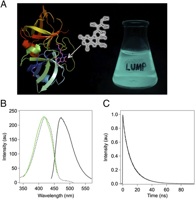
Fig. 1.
Structure and fluorescence properties of LUMP. (A) Crystal structure of LUMP (8) with surface-bound ribityl-lumazine adjoined with a photograph of a flask containing an E. coli culture expressing LUMP. (B) Peak-normalized absorption (dashed line), excitation spectrum (green line) with emission at 470 nm, and emission spectrum (solid line) of LUMP measured in aqueous buffer [20 mM Hepes (pH 7.9), 150 mM NaCl] at 20 °C. (C) Time-resolved fluorescence intensity decay of LUMP. au, arbitrary units.

