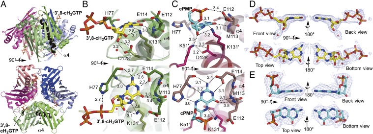
Fig. 2.
Crystal structure of MoaC in complex with 3′,8-cH2GTP or cPMP. (A) The hexameric structure (a trimer of homodimers) of K51A-MoaC with 3′,8-cH2GTP (shown in sticks) bound at the N terminus of helix α4 (highlighted in black). (B and C) The active sites of K51A-MoaC in complex with 3′,8-cH2GTP (B) and WT-MoaC with cPMP (C). Ligands (3′,8-cH2GTP in yellow; cPMP in blue) and the catalytically essential residues are shown as sticks. H-bond interactions are indicated by dashed lines with distances in angstroms. (D and E) Simulated annealing Fo−Fc electron density maps (mesh, contoured at 3 σ). Shown is the electron density in the active-sites of K51A-MoaC•3′,8-cH2GTP (D) and WT-MoaC•cPMP (E).

