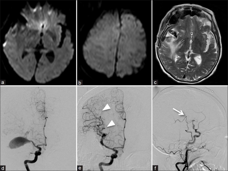
Figure 2.

(a and b) Axial diffusion-weighted MR images obtained 1 day after surgery revealing no acute ischemia. (c) Axial T2-weighted MR image performed 1 day after surgery showing no new changes other than the preexisting edema around the aneurysm. (d) Preoperative angiogram demonstrating a giant MCA aneurysm. (e) Postoperative angiogram showing the complete obliteration of the aneurysm with preservation of the parent artery. Note the remarkable increase of the arterial flow in the MCA territory (arrowheads). (f) Right lateral carotid angiogram demonstrating that the bypass flow covered only a small area of the frontal lobe distal to the site of anastomosis (arrow)
