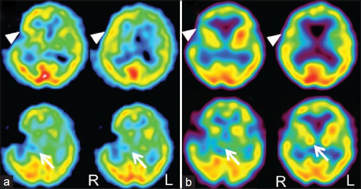
Figure 3.

(a) 99mTc-ECD SPECT performed 3 days after surgery revealing hyperperfusion in the frontal cortex (arrowheads). There was also slight hypoperfusion in the right basal ganglia including the subthalamic nucleus (arrows). (b) 99mTc-ECD SPECT obtained 8 weeks after surgery showed the resolution of hyperperfusion in the right frontal cortex (arrowheads) with the resolved laterality of the perfusion in the subthalamic regions (arrows)
