Figure 2.

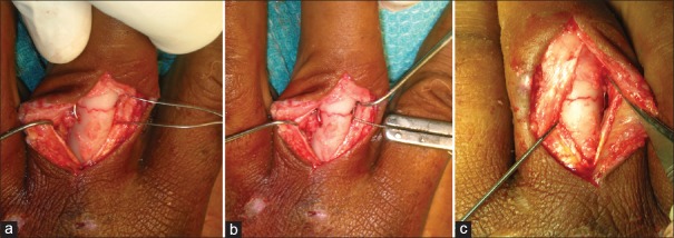
Intraoperative clinical photographs of theta fixation. (a) Fracture exposed through the dorsal extensor splitting approach, reduced and cerclage wire inserted. (b) Cerclage wire tightened. (c) Kirschner-wire inserted

Intraoperative clinical photographs of theta fixation. (a) Fracture exposed through the dorsal extensor splitting approach, reduced and cerclage wire inserted. (b) Cerclage wire tightened. (c) Kirschner-wire inserted