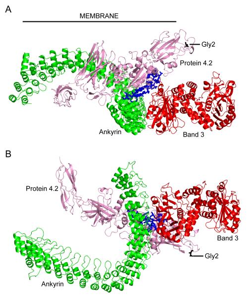
Figure 2. Ankyrin-band 3-protein 4.2 complex.
(A) view from within the plane of the membrane, approximate location of the inner face of the cell membrane is indicated with a black line; (B) view from the cytoplasm. Proteins are displayed as Cα cartoon, ANK repeat region of ankyrin in green, cytoplasmic domain of band 3 (disordered regions omitted) in red and protein 4.2 in pink. Protein 4.2 residues 187-211 (associated with ankyrin and band 3 binding) are coloured blue, and displayed as sticks; residue 2 (myristolyation site) is coloured black and displayed as sticks. For interpretation of the references to colour in this figure legend, the reader is referred to the web version of this article.

