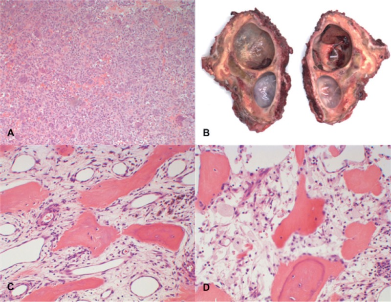
Figure 3.
A. Histology of the biopsy specimen before the start of denosumab treatment showed numerous uniformly spaced giant cells with large vesicular nuclei, prominent nucleoli, and mononuclear cells with eosinophilic cytoplasm and nuclei similar to those of adjacent giant cells, which is consistent with the diagnosis of GCTB. B. Macro specimen of intralesional resection after 7 months of treatment with denosumab, showing 2 large cysts and focal clots. The resection planes were not free of reactive tissue. C and D. Histology of the surgical specimen showed stromal cells, scattered mononuclear spindle cells with elongated oval-shaped nuclei without evident atypia, and diffuse foamy macrophages. No multinucleated giant cells were seen.

