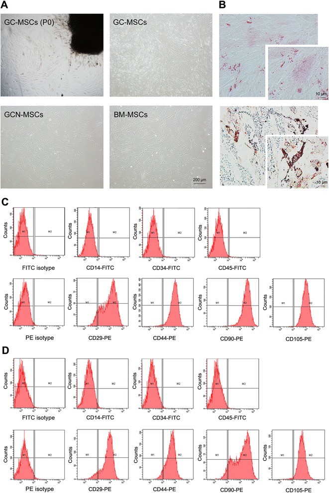
Fig. 1.

Characterization of human gastric cancer-derived MSCs. (A) Spindle-shaped cells migrated from gastric cancer tissues after 7–10 days of primary culture (upper left) and fibroblast-like cells appeared at passage 4 of GC-MSCs culture (upper right) with the morphology similar to GCN-MSCs (lower left) or BM-MSCs (lower right) (×40). (B) Representative photographs of GC-MSCs differentiated into mineralizing cells with alizarin red S staining (upper) and adipogenic cells with Oil red O staining (lower) (×200, ×400). (C) Surface antigens expressed on GC-MSCs analyzed by flow cytometry. (D) Surface antigens expressed on GCN-MSCs analyzed by flow cytometry
