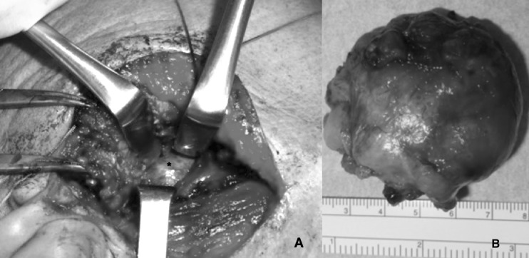
Fig. 1.
(A) tumour of the deep lobe (asterisk) under the superficial parenchyma, removed performing an ECD after TPA (a portion arising from the deep tumour occupies part of the superficial lobe and is surrounded by some branches of the facial nerve); (B) the tumour (pleomorphic adenoma) after the procedure.

