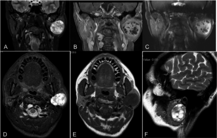
Fig. 1.
MRI images showing left parotid lesion with extension in the deep parotid lobe close to the stylomastoid process (as appears in the axial, coronal and sagittal plane). The lesion appears on hyperintense fat-suppressed T2-weighted images (T2WI) and hypointense on T1-weighted images (T1WI), with marked enhancement on gadolinium-enhanced T1WI with cystic changes inside the lesion. All these features are similar to those observed in cases of pleomorphic adenoma.

