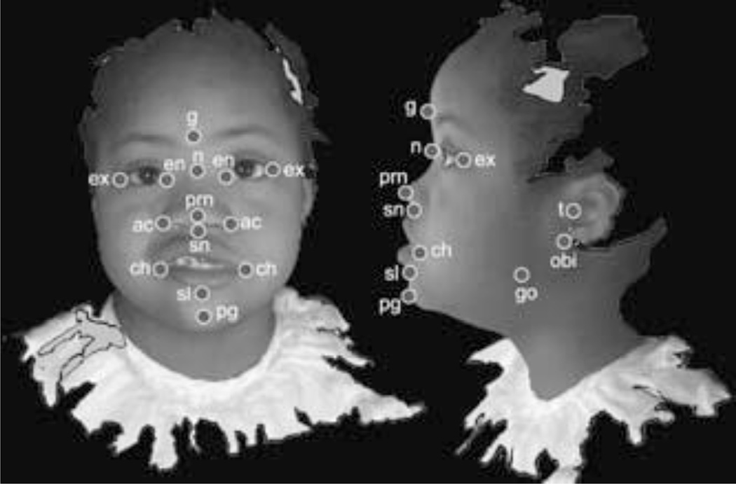
Figure 2.
Landmarks collected from 3dMD images. Landmarks are illustrated on 2D facial and lateral views taken from screenshots of an example 3dMD image, though actual data are 3D. Glabella (g), Nasion (n), Pronasale (prn), Subnasale (sn), Sublabiale (sl), Pogonion (pg), Endocanthion left and right (en), Exocanthion left and right (ex), Alar curvature (ac), Chelion (ch), Tragion (t), Otobasion inferius (obi), Gonion (go).

