
Figure 2. Example of benign and malignant nodules in a single patient.
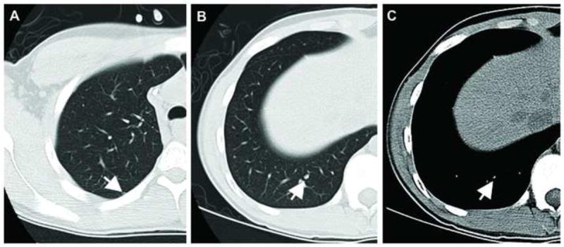
Axial CT images from a 17-year-old male patient with osteosarcoma. Image (A) shows a pathologically proven benign nodule which was rated by all three readers as either indeterminate or benign. The calcified nodule in (B) and (C) (lung and mediastinal window settings, respectively) was rated by all three readers as malignant and proved to be malignant at resection.