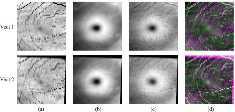
Figure 2.
Fundus registration of 2 visits. Columns (a), (b), and (c) show the vessel shadow projection images S, the retina thickness images T , and the combined FPIs, respectively. In (d), we show color overlay images (top) before and (bottom) after registration. White indicates the improved alignment of the vessels.

