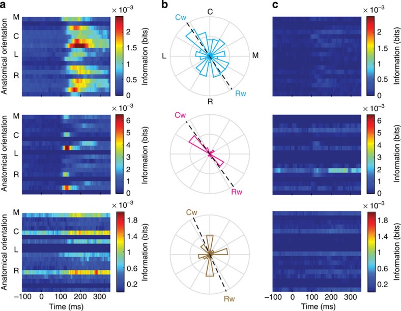
Figure 7. Target-direction information content of spike sequencing between neurons with narrow spike waveforms.
Data from monkeys Rs (top), Mk (middle) and Rj (bottom). (a) Time-resolved, mutual information between spike sequencing and target direction (that is, the direction from the location of the acquired target to the new target). Information was calculated for only pairs of neurons with excitatory connections. The colour map consists of 18 rows corresponding to 18 orientations (20-degree bins) aligned to anatomical orientations denoted with M, medial; C, caudal; L, lateral and R, rostral, with information in bits in blue-red false colour. (b) Circular distributions of spike sequencing information with respect to the orientation of neuron pairs on the cortical surface. Spike sequencing information was summed over the time window in [100, 350] ms and over pairs within each orientation and then normalized. The dashed black lines represent the beta wave axis defined by Cw (caudal wave direction) and Rw (rostral wave direction) as in Fig. 1d. (c) Target-direction information content of sequencing firing of non-connected narrow-spiking neurons.

